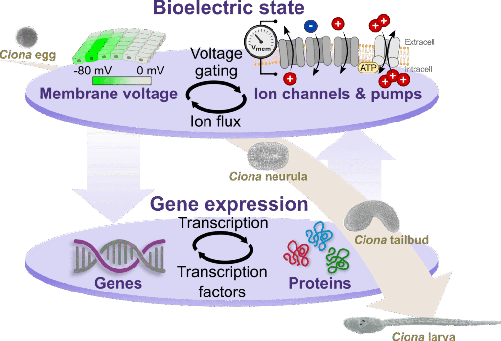
Bioelectricity and gene regulation interplay in embryonic development
Embryonic development is a closely regulated process influenced by multiple biological signals, such as chemical gradients and gene regulation. Recent research has identified bioelectric signals, produced by the movement of ions across cell membranes, as another component involved in this process. The precise mechanisms through which these bioelectric signals interact with gene expression remain under investigation.
The ELECTROGENDEV project aims to examine the interaction between bioelectric signals and gene expression. Using ascidians—marine invertebrates related to vertebrates—the study will map the bioelectric landscape and determine the ion channels involved in generating these signals. By identifying points of convergence between bioelectric activity and gene regulation, the research seeks to establish a comprehensive model of embryonic development, with potential implications for regenerative medicine and developmental disease treatment.
This project is led by Josep MARTÍ SOLANS and is funded by the European Union under Marie Skłodowska-Curie Actions (MSCA).
AscidianPnsEvo (2024-2028)
Comparative study of peripheral nervous system formation in ascidians: conservation, drift and variability.
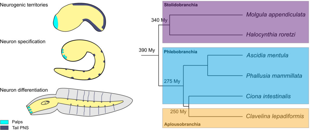
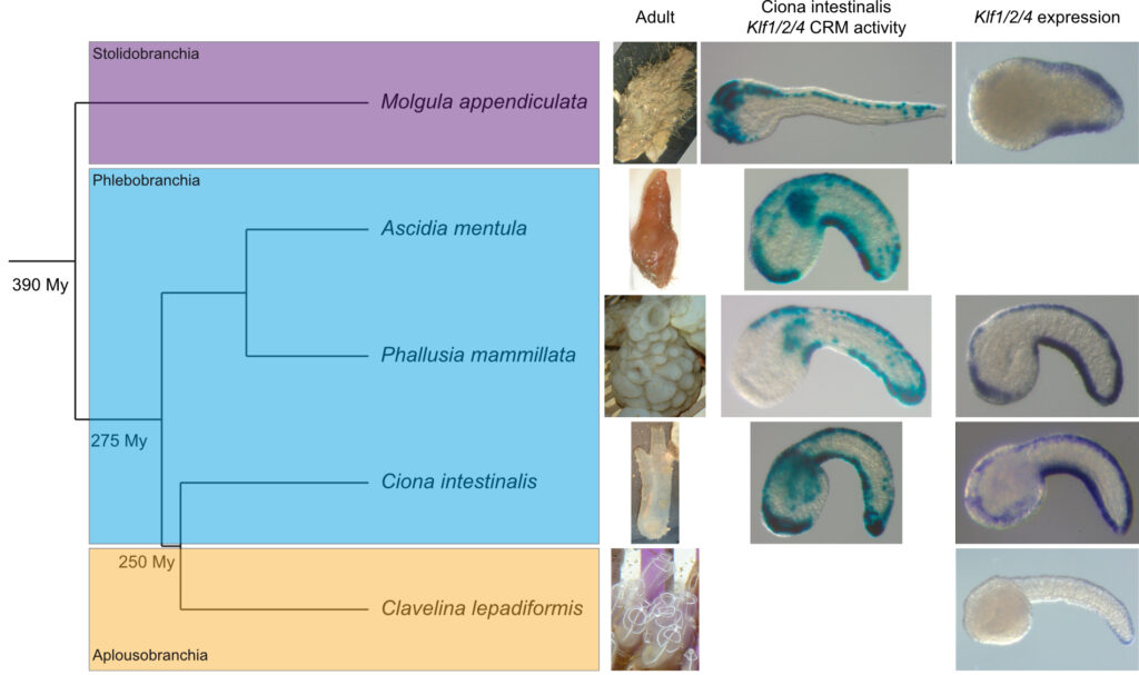
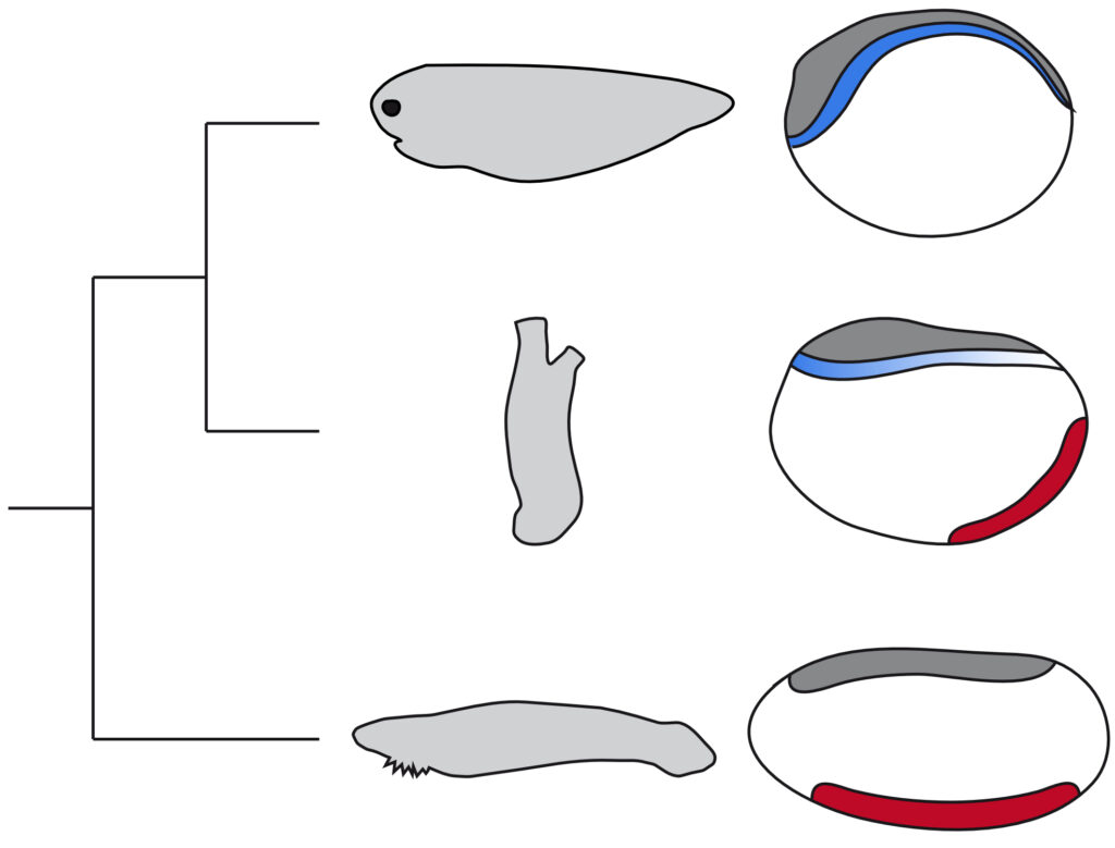
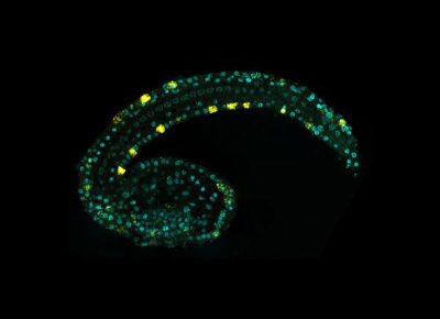
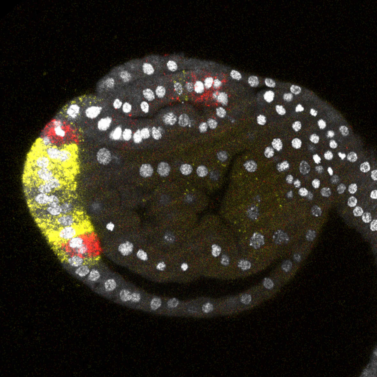
In vertebrates, the peripheral nervous system (PNS) originates from transient embryonic structures, the neural crest and the placodes. Since these cell populations also give rise to a variety of cell types and structures that make vertebrates unique, understanding their formation and identifying their evolutionary origins has been the focus of intense study. Ascidians, marine invertebrates that belong to the vertebrate sister group, do not have bona fide neural crest or placodes. But their simple PNS made of a small number of epidermal sensory neurons display a number of similarities with the vertebrate PNS: topological organization, specification mechanisms and gene expression profiles. However, a proper comparison between ascidians and vertebrates is impaired by high levels of developmental system drift in ascidians, a fast evolving lineage. Aiming at defining the ancestral state of the ascidian PNS and the underlying developmental mechanisms is thus crucial for understanding the emergence of the vertebrate PNS. To achieve this goal, we propose a close functional comparative approach of PNS formation in different ascidian species. We will characterize the PNS and determine how the gene regulatory networks deciphered in the reference species Ciona intestinalis are deployed. We will focus on gene expression and its regulation by examining the function of inputs from signaling pathways, and the activity and evolution of cis-regulatory elements. We should be able to propose an ancestral PNS network and describe changes that took place during ascidian diversification. In addition, we will concentrate on two parts of the PNS: the anterior palps that have a fixed number of neurons in different species, and the tail PNS with a variable number between individuals of a given species, and between species. This will enable to test the hypothesis that the variability of a trait is linked to its evolvability and to the divergence/drift of its regulatory mechanisms.
This project is funded by the ANR.
AnimalCellulose (2021-2023)
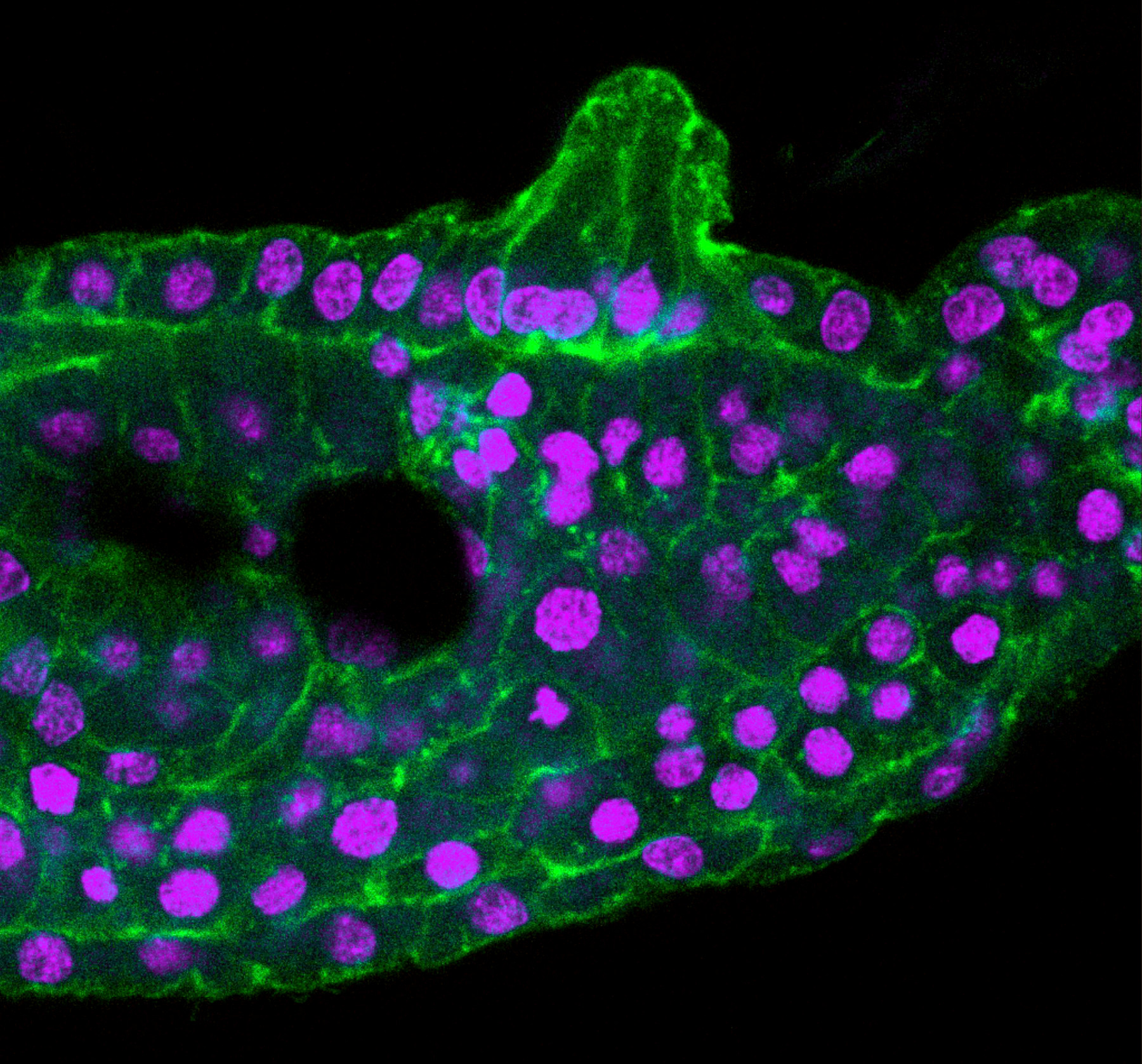
Tunicates are characterized by a unique feature: they are the only animals that produce cellulose. This capacity, restricted to plants, fungi and bacteria, has appeared through the acquisition of a cellulose synthase gene by horizontal transfer. Cellulose is key in ascidians since it is the major component of the tunic, an acellular exoskeleton-like structure secreted by the epidermis that surrounds the animal both in the adult and in the tadpole larva. The goal of our project is to use the knowledge on molecular patterning of the epidermis during embryogenesis as an entry point to uncover mechanisms controlling tunic production and 3D morphogenesis by extracellular material.
This project is financed by Sorbonne Université within the frame of the Emergence program.
Our team belonging to a marine station, we ambition to establish novel ascidian species as models for experimental molecular embryology. We aim at comparing, with the reference species Ciona intestinalis, the cellular and molecular mechanisms regulating embryonic development. We are working at generating resources (transcriptomes, genomes…), methods (in situ hybridization, electroporation…), and data (expression patterns, cis-regulatory elements activity…) for several local species.
This project is supported by the CNRS (INSB) in the frame of the Diversity of Biological Mechanisms call.
VentralPNS (2018-2021)
While vertebrate peripheral nervous system (PNS) exclusively arises from dorsal structures (neural crest and placodes), a large part of invertebrate chordate (ascidians and amphioxus) PNS originates from the ventral ectoderm. BMP signals induce a neurogenic field from which sensory neurons are selected by Notch signaling. This ventral PNS (vPNS) likely corresponds to an ancestral chordate structure that may have been lost in vertebrates or shifted dorsally to form placodes/neural crest, possibly via co-option of ancestral specification mechanisms. Our project aims at exploring functionally the ventral PNS using both groups of invertebrate chordates as references. We will perform a thorough side-by-side comparison to 1) identify vPNS genes, 2) define their hierarchy and transcriptional regulation, 3) determine whether their mode of regulation is conserved and 4) probe the existence of vertebrate counterparts for either a vPNS territory, vPNS neurons or vPNS gene regulatory networks.
This ANR funded project is in collaboration with two other teams from the BIOM:
– Group E1 Evolution and Development of Chordates led by Hector ESCRIVA and Stéphanie BERTRAND (amphioxus)
– Group E3 Development and evolution of vertebrates led by Sylvie MAZAN (catshark and lamprey)
ChorMedFin (2012-2015)
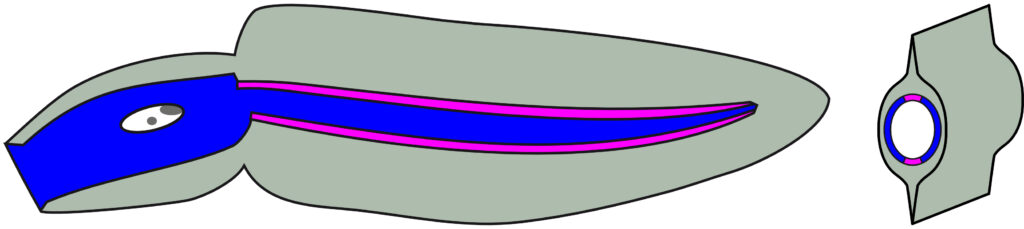
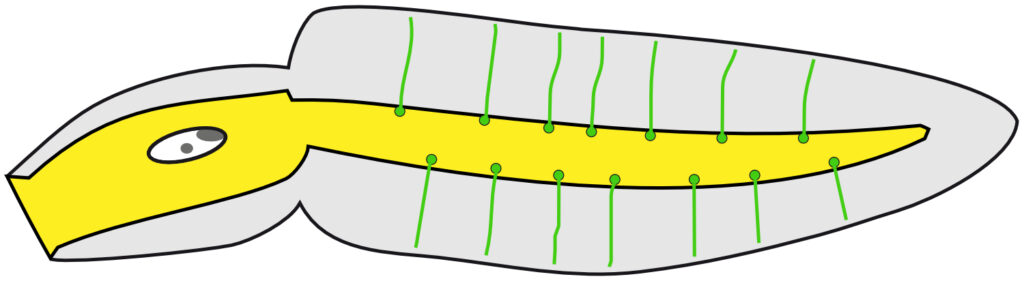
Ciona larval tail epidermis is characterized by a crystal-like cellular organization of only 8 rows of cells, each endowed with a specific expression profile. We have uncovered key events that establish this patterning and control the formation of two derivatives along the midlines: the peripheral nervous system (composed of epidermal sensory neurons) and the median fin. This latter structure is a fascinating case of morphogenesis without cells. The tunic surrounding the larva is formed by the secretion of extracellular material (including cellulose) extending along the ventral and dorsal midlines to form the fin blades. We have proposed that the tail epidermal neurons and the fin may have common origins with vertebrate structures, the neural crest and/or placodes and the median fin, respectively.
In order to get insights into the formation and evolutionary history of the median fin and peripheral nervous system in chordates, we propose to:
1) Reconstruct the GRN in Ciona.
2) Describe the development of the equivalent structures and uncover their specification in other basal chordates.
3) Test GRN conservation.
The ChorMedFin (Chordates Median Fin: Gene regulatory networks controlling median fin formation in basal chordates) project has been funded by the ANR.